








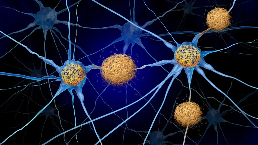
Protecting brain cells from destruction remains one of the most formidable challenges in the field of neurology. For decades, researchers have grappled with the mechanisms underlying Alzheimer’s disease and a group of related neurodegenerative conditions collectively known as tauopathies. These disorders are characterized by the abnormal accumulation of "tau" protein tangles within the brain, a process that invariably leads to the death of nearby neurons. As these tangles proliferate, patients experience a progressive loss of memory, cognitive function, and, eventually, their independence.
However, a paradigm shift is currently underway in the scientific community. While tau tangles have long been the primary target for therapeutic intervention, growing evidence suggests that the protein itself may not be the sole culprit in the widespread neuronal death observed in these diseases. Instead, a new, compelling narrative is emerging: the brain’s own immune system, when triggered by the presence of tau, may be the true architect of the damage. By identifying a way to interrupt this misguided immune response, researchers at Washington University School of Medicine in St. Louis have uncovered a promising, novel strategy to preserve brain health.
The Role of T Cells in Brain Damage
The study, published September 28 in the journal Neuron, highlights a critical discovery regarding how immune cells, specifically T cells, infiltrate the brain in response to tau accumulation. In experiments involving mice genetically predisposed to develop tau deposits—mirroring the pathology of human Alzheimer’s—researchers focused on a protein called CXCR3. This protein acts as a beacon, guiding T cells into the brain tissue. By administering an antibody designed to block CXCR3, the research team successfully reduced the number of T cells inside the brains of these mice by approximately 50 percent over a period of several months.
The results of this intervention were striking. The treated mice not only retained roughly 40% more brain tissue in critical memory-related regions compared to their untreated counterparts, but they also demonstrated superior performance on standardized memory tests. Perhaps most significantly, these protective benefits were achieved without altering the amount of tau in the brain. This finding suggests that it is the immune system’s aggressive reaction to the protein, rather than the tau tangles themselves, that drives the most catastrophic loss of neurons and cognitive function.
"In tauopathies, including Alzheimer’s disease, there’s no treatment right now that actually decreases neurodegeneration," said David M. Holtzman, MD, senior author of the study and the Barbara Burton and Reuben M. Morriss III Distinguished Professor in the Department of Neurology at WashU Medicine. "If we can show that we’re really decreasing brain cell death, it’s certainly worth trying to pursue that pathway to prevent the most devastating consequences of these diseases."
Beyond Amyloid: Reframing Alzheimer’s Pathology
To understand the significance of this discovery, it is necessary to consider the current landscape of Alzheimer’s treatment. For years, the scientific focus has been heavily tilted toward the removal of amyloid plaques—deposits that form between brain cells in the early stages of the disease. Recent FDA-approved medications, such as lecanemab and donanemab, represent major breakthroughs in this area, as they effectively target these plaques to slow cognitive decline.
However, these therapies have significant limitations. While they can delay the progression of symptoms, they have not been shown to prevent the death of brain cells. Furthermore, they are ineffective for primary tauopathies—conditions where tau buildup occurs independently of amyloid. Alzheimer’s disease is classified as a secondary tauopathy because it involves both amyloid plaques and tau tangles. The fact that current treatments target only one of these two pathologies highlights an urgent need for therapies that address the downstream damage caused by tau.
Dr. Holtzman’s laboratory has spent years investigating the immune system’s role in this process. A 2023 study published in Nature laid the groundwork for the current findings, revealing that T cells indeed migrate into the brains of mice with tau accumulation, specifically congregating in regions where the protein density is highest. A separate, related study published earlier this month in Nature Neuroscience provided another piece of the puzzle, confirming that the chemical signals directing these T cells are generated in the lymph nodes outside the brain.
Navigating the Pathway: The CXCL10-CXCR3 Axis
The process by which T cells locate and infiltrate the brain is a complex biological journey. T cells move through the body by tracking chemical signals known as chemokines. Previous research in Holtzman’s laboratory had identified elevated levels of a specific signal called CXCL10 in the brains of mice with tau buildup, a finding that correlates with elevated CXCL10 levels observed in human patients with Alzheimer’s.
Activated T cells express a receptor on their surface called CXCR3, which functions as a "lock" that recognizes and binds to the "key" provided by CXCL10. This pairing forms the CXCL10-CXCR3 pathway, which the researchers hypothesized was the primary mechanism guiding inflammatory T cells into the brain. To test this, the team, which included co-senior author Jason Ulrich, PhD, and lead author Joshua T. Emmerson, PhD, studied mice lacking either CXCL10 or the CXCR3 receptor. In both scenarios, the researchers observed that T cells were completely unable to infiltrate the brain, even when the researchers intentionally induced systemic inflammation to provoke an immune response.
This confirmed that the CXCL10-CXCR3 interaction is the essential gateway for these harmful immune cells. By demonstrating that the pathway is both necessary and targetable, the researchers established a clear mechanism for potential therapeutic intervention.
A New Frontier for Drug Development
The experimental treatment used in the study—a monoclonal antibody—holds profound implications for the future of drug design. One of the greatest hurdles in treating neurological disorders is the blood-brain barrier, a selective, semi-permeable border that prevents many substances from entering the brain from the bloodstream. Historically, developers have had to design drugs capable of crossing this barrier, a process that is often difficult, costly, and inefficient.
In this study, the researchers observed that the antibody did not actually enter the brain tissue; it stopped at the boundary. Yet, it was still highly effective at reducing neurodegeneration. This suggests that scientists may not need to design drugs that penetrate the brain to achieve a therapeutic effect. By targeting the immune response at the periphery—such as in the lymph nodes or the bloodstream—it may be possible to stop the inflammatory cascade before it ever reaches the brain.
"For this therapeutic approach, if it is safe, you wouldn’t have to design the drug to get into the brain—which is a big deal since most molecules don’t cross the blood-brain barrier well—and you don’t have to get rid of the tau to get this therapeutic effect," Dr. Holtzman noted.
Moving Toward Clinical Application
While the results in mice are highly encouraging, the research team remains cautious, noting that the study is a foundational step. Further research is required to determine the safety and long-term efficacy of this approach in humans. However, the path forward may be shorter than anticipated. Because drugs that modulate T cells are already utilized to treat other conditions—such as multiple sclerosis and various autoimmune diseases—researchers may be able to repurpose existing therapies to investigate their potential in treating Alzheimer’s and other tauopathies.
This shift in strategy represents a fundamental change in how the medical community views neurodegeneration. Rather than viewing tauopathies purely as protein-misfolding disorders, this research suggests they should also be considered in the context of immune-mediated damage.
"Tauopathies aren’t thought of as autoimmune disorders, so they haven’t been treated the same way, but this study shows for the first time in an animal model that these diseases respond to a specific T-cell therapy," said Dr. Holtzman, who also directs the Hope Center for Neurological Disorders and the Knight Alzheimer Disease Research Center at WashU Medicine.
By moving the focus from the buildup of proteins to the immune system’s reaction to those proteins, scientists may be opening a new door to effective, disease-modifying treatments. If successful in human trials, this approach could offer a way to preserve brain tissue and cognitive function, providing a beacon of hope for the millions of people currently living with or at risk for Alzheimer’s and related diseases. The research underscores the necessity of interdisciplinary collaboration, combining immunology, neurology, and drug development to solve one of the most pressing health challenges of our time.
The study received broad support from organizations including the National Institutes of Health, the BrightFocus Foundation, the GHR Foundation, the Carol and Gene Ludwig Initiative in Neuroimmunology Research, and several private donors. Dr. Holtzman holds multiple disclosures related to his work, including co-founding C2N Diagnostics and serving on the scientific advisory boards of several major pharmaceutical and biotech firms, ensuring his research remains at the cutting edge of clinical application.