







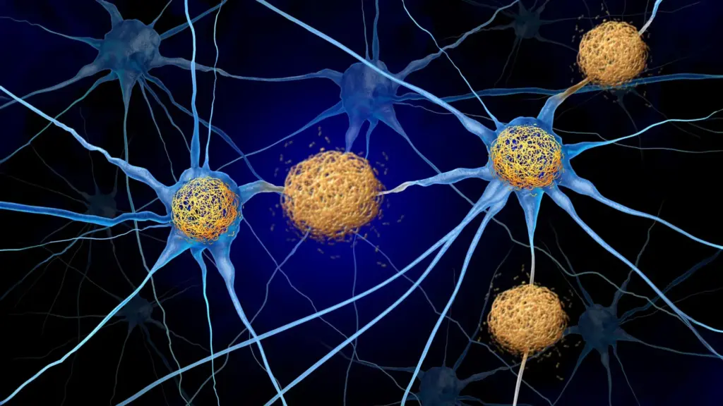
Protecting brain cells from destruction remains one of the most formidable challenges in the field of neurology. For decades, the primary focus of Alzheimer’s disease research and the study of related neurodegenerative conditions—collectively known as tauopathies—has been the accumulation of abnormal protein tangles. These tangles, composed of a protein called tau, build up inside the brain, and as they accumulate, nearby neurons inevitably begin to perish. However, a significant shift in perspective is currently underway. Growing evidence suggests that the tau protein itself may not be the primary culprit behind the widespread devastation of brain cells, memory, and cognitive independence. Instead, it is the immune system’s misdirected reaction to these protein deposits that appears to be the engine driving the catastrophic loss of neuronal tissue.
In a landmark study published September 28 in the journal Neuron, scientists at the Washington University School of Medicine in St. Louis have identified a promising, novel strategy for interrupting this damaging immune response. By targeting the pathway that allows specific immune cells to infiltrate the brain, researchers have successfully preserved memory and brain tissue in animal models, even while the underlying tau tangles remained untouched.
The Role of T Cells in Neurodegeneration
The study, led by senior author David M. Holtzman, MD, the Barbara Burton and Reuben M. Morriss III Distinguished Professor in the Department of Neurology at WashU Medicine, sought to understand the mechanics of neurodegeneration in tauopathies. Currently, there is no treatment that effectively halts or reverses the death of brain cells in these disorders. "In tauopathies, including Alzheimer’s disease, there’s no treatment right now that actually decreases neurodegeneration," Holtzman explained. "If we can show that we’re really decreasing brain cell death, it’s certainly worth trying to pursue that pathway to prevent the most devastating consequences of these diseases."

Current clinical approaches to Alzheimer’s, such as the FDA-approved medications lecanemab and donanemab, are primarily designed to clear amyloid plaques—deposits that form between brain cells during the early stages of the disease. While these therapies have proven capable of slowing cognitive decline by addressing the initial amyloid cascade, they have not demonstrated the ability to prevent the terminal death of neurons. Furthermore, they offer no relief for primary tauopathies, where tau builds up independently of amyloid. Because Alzheimer’s involves both amyloid and tau, it is classified as a secondary tauopathy, but the persistence of tau-driven damage remains a critical unmet medical need.
The Holtzman laboratory has long hypothesized that the immune system might hold the key to unlocking this mystery. Their investigations, including a 2023 study published in Nature, confirmed that T cells—a specialized type of immune cell—do indeed penetrate the brains of mice suffering from abnormal tau accumulation. These cells tend to congregate in the exact brain regions where tau deposits are most dense, effectively turning those areas into sites of chronic inflammation and neuronal destruction. A subsequent study, published earlier this month in Nature Neuroscience, provided a critical piece of the puzzle: the signals directing these T cells into the brain originate from lymph nodes located outside the central nervous system.
Identifying the Chemical "Roadmap"
To halt the infiltration of these harmful T cells, researchers needed to understand the "roadmap" they use to navigate the body. T cells follow chemical gradients, specifically proteins known as chemokines, to reach their destination. Through previous experiments, the team identified that mice with tau accumulation exhibited unusually high levels of a chemokine called CXCL10. This finding is particularly significant because clinical data has also shown elevated levels of CXCL10 in the brains of human Alzheimer’s patients.
Activated T cells possess a surface receptor called CXCR3, which functions like a molecular antenna, allowing the cells to "lock on" to CXCL10. This discovery suggested a clear pathway: the CXCL10-CXCR3 axis serves as the chemical beacon guiding inflammatory T cells into the brain. To validate this, the research team—including co-senior author Jason Ulrich, PhD, a professor of neurology at WashU Medicine, and first author Joshua T. Emmerson, PhD, a postdoctoral researcher—conducted experiments with mice that were genetically engineered to develop tau deposits.
The results were unequivocal. When the mice lacked either the CXCL10 protein or the CXCR3 receptor, the T cells were unable to infiltrate the brain. Even when the researchers introduced external inflammation to provoke an immune response, the brains remained protected from T-cell infiltration, demonstrating that the CXCR3-CXCL10 signaling pathway is the primary gatekeeper for these immune cells.
Preserving Memory and Brain Tissue
With the mechanism identified, the team transitioned to a therapeutic intervention. They selected young mice that had already begun to develop tau deposits but had not yet reached the stage of widespread neuronal death. For a period of three and a half months, the researchers administered an antibody designed to block the CXCR3 receptor once every five days.
The outcomes were striking. By the conclusion of the study, the treated mice had roughly half the number of T cells in their brains compared to the untreated control group. More importantly, this reduction in immune infiltration translated into tangible physical and cognitive benefits. The treated mice exhibited significantly less evidence of neuronal injury and retained approximately 40% more tissue in key brain regions associated with memory and cognitive function. On behavioral memory tests, the treated mice consistently outperformed their untreated counterparts.
Perhaps the most surprising finding was that these neuroprotective effects occurred despite the fact that the total amount of tau tangles remained unchanged. The treated and untreated mice had similar levels of protein accumulation, yet the treated mice showed substantially less brain damage. This suggests that the destruction of brain tissue is not an inevitable consequence of tau accumulation itself, but rather a consequence of how the immune system chooses to react to that tau. By dampening the immune system’s "overreaction," researchers can protect the brain even if the offending protein cannot be immediately cleared.
A New Frontier for Therapeutic Development
The implications of this research for future drug development are profound. One of the most significant barriers in treating neurological diseases is the blood-brain barrier, a tightly packed layer of cells that protects the brain from harmful substances in the blood but also prevents the vast majority of therapeutic drugs from entering the brain tissue.
In this study, the antibody used to block CXCR3 reached the boundary of the brain but did not need to cross into the brain tissue itself to be effective. Because the signal directing the T cells originates outside the brain—in the lymph nodes—the treatment works by intercepting the immune cells before they ever reach the central nervous system. "For this therapeutic approach, if it is safe, you wouldn’t have to design the drug to get into the brain—which is a big deal since most molecules don’t cross the blood-brain barrier well—and you don’t have to get rid of the tau to get this therapeutic effect," Dr. Holtzman noted.
While these results are highly encouraging, the research remains in the preclinical phase, having been tested exclusively in mouse models. The transition to human clinical trials will require extensive safety and efficacy testing. However, the path forward may be shorter than anticipated. Dr. Holtzman suggested that researchers might look to existing, approved medications used to manage T cells in other diseases, such as multiple sclerosis or other autoimmune conditions, to see if they could be repurposed to mitigate Alzheimer’s-related brain damage.
This would represent a fundamental shift in the treatment landscape for neurodegenerative disorders. Rather than focusing solely on the clearance of abnormal proteins—a task that has proven notoriously difficult—medical science may move toward a dual strategy: managing the proteins while simultaneously "calming" the immune system to prevent the collateral damage that leads to cognitive decline.
"Tauopathies aren’t thought of as autoimmune disorders, so they haven’t been treated the same way, but this study shows for the first time in an animal model that these diseases respond to a specific T-cell therapy," said Holtzman, who also directs the Hope Center for Neurological Disorders and the Knight Alzheimer Disease Research Center at WashU Medicine. By refocusing efforts on the intersection of immunology and neurology, researchers hope to move closer to a time when Alzheimer’s can be managed not just by addressing its deposits, but by protecting the integrity of the brain from the inside out.
The research was made possible through the support of the National Institutes of Health and the National Institute on Aging, alongside contributions from the BrightFocus Foundation, the GHR Foundation, the Carol and Gene Ludwig Initiative in Neuroimmunology Research, and various private donors, including the Freedom Together Foundation. As the scientific community digests these findings, the focus will now shift to identifying how this pathway can be safely modulated in humans, potentially opening a new door in the fight against the world’s most pervasive neurodegenerative diseases.