






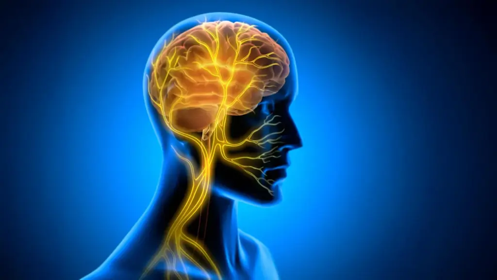
Why do some new skills seem to click almost instantly, becoming second nature after only a few attempts, while others remain frustratingly elusive despite hours of dedicated practice? For decades, the consensus among neuroscientists and educators has been that the difference lies in a combination of innate talent, the intensity of focus, and the sheer volume of repetition. However, new research suggests that the secret to mastery may involve an overlooked biological factor: the condition of the brain’s internal environment during the post-practice period. A study conducted by researchers at Tohoku University indicates that the body’s communication with the brain, mediated through the vagus nerve, may act as a critical gatekeeper for turning fleeting practice into lasting learning.
The findings, published in iScience on August 25, 2026, suggest that learning is not a process that occurs in isolation within the cranium. Instead, it is an integrated bodily function, heavily influenced by the constant stream of information flowing from internal organs to the brain. By utilizing vagus nerve stimulation (VNS) in a controlled setting, the research team has demonstrated that the timing of this neural signaling can significantly strengthen motor learning, potentially uncovering a hidden mechanism that helps new skills persist in the long term.
The Vagus Nerve as a Learning Conduit
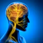
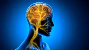
The vagus nerve is often described as one of the most vital information highways in the human body. As a key component of the autonomic nervous system, it facilitates a continuous, bidirectional dialogue between the brain and the body’s internal organs, such as the heart, lungs, and digestive tract. While the brain sends "top-down" instructions to these organs to regulate physiological functions, the vagus nerve simultaneously carries "bottom-up" sensory information back to the brain, providing a real-time status update of the body’s internal state.

Scientists have long been interested in the potential to manipulate this pathway. Vagus nerve stimulation (VNS) is a clinically approved intervention already used to treat various medical conditions, including epilepsy and treatment-resistant depression. Traditionally, the therapeutic efficacy of VNS has been attributed to its ability to modulate neurotransmitter systems—the chemical messengers that dictate how neurons communicate with one another. However, the team at Tohoku University’s super network brain physiology lab suspected that this was only part of the story. Their investigation points toward a previously underappreciated mechanism: the regulation of rhythmic blood flow within the brain.
To test this hypothesis, the researchers performed a series of experiments on mice. They implanted a specialized, miniaturized cuff electrode around the left cervical vagus nerve. This allowed the team to deliver precise electrical stimulation at specific intervals. The mice were then trained on a horizontal optokinetic response (HOKR) task. This task is a well-established model for studying cerebellar-dependent motor learning, requiring the mice to improve their ability to track moving visual stripes. The movement is analogous to the subconscious stabilization of the eyes a human experiences when standing on a train platform and watching a locomotive pull away, an automatic reflex that must be fine-tuned through neural plasticity.
The Crucial Window: Post-Practice Stimulation
One of the most significant aspects of the study was the timing of the intervention. The researchers chose to apply VNS strictly after the completion of each training session, rather than during the learning task itself. This decision was based on the premise that the consolidation of memory—the process by which a temporary, fragile memory trace is stabilized into a durable, long-term structure—occurs primarily during the periods following active practice.
The results were striking. The stimulation provided no immediate, visible improvement in performance while the mice were actively engaged in the task. Instead, the benefits emerged in the days that followed. Mice that received the post-training VNS demonstrated significantly more robust, long-term learning compared to control groups that did not receive the stimulation. This outcome strongly supports the theory that VNS does not necessarily make the brain "smarter" in the moment, but rather optimizes the neurological landscape for the stabilization of new information.
Professor Ko Matsui, a lead investigator on the project, emphasizes the significance of this timing. "The key point is that VNS was delivered only after training," Matsui explains. "Our findings suggest that VNS may open a hidden window of opportunity for enhanced learning by making the brain environment more receptive to long-lasting change." By intervening during this post-training consolidation phase, the researchers were able to effectively prime the brain to lock in the motor patterns that had been practiced, turning transient neural activity into enduring memory.
Rhythmic Vascular Responses and Brain Plasticity
To uncover the physiological basis for this enhanced learning, the team utilized fiber photometry to observe real-time changes within the brain. They focused their measurements on the cerebellar flocculus, a specific area of the cerebellum that is critical for the coordination of eye movements and the acquisition of the HOKR task.
The researchers observed that a single round of VNS triggered a distinct, two-phase vascular response. Initially, there was a brief, localized decrease in blood volume, followed by a delayed rise. When the researchers repeated the stimulation, they observed the creation of rhythmic oscillations in blood volume—a pulsing effect that appeared to mirror the timing of the VNS.
Crucially, these vascular rhythms were not merely incidental. The team found a direct correlation between the magnitude of these blood volume oscillations and the success of the learning task. Mice that exhibited larger, more consistent vascular rhythms during the post-training window consistently showed superior performance by the fifth day of the experiment. This discovery suggests that the brain’s vascular environment—its metabolic supply chain—is a dynamic partner in the learning process. By influencing the rhythmic flow of blood, VNS may be optimizing the delivery of oxygen and nutrients, or perhaps facilitating the clearance of metabolic byproducts, thereby creating the ideal conditions for synaptic remodeling.
Lead author Junyu Chen highlights the implications of these findings, noting that the brain is far more responsive to the body’s internal state than current paradigms might suggest. "Our brains may be more strongly influenced by the body than we imagine," says Chen. "By tuning the brain’s metabolic environment, including rhythmic vascular movements, we may eventually unlock capacities that would otherwise remain latent." This suggests that the body is not merely a vessel for the brain but an active participant in defining the limits of cognitive and motor potential.
Expanding the Frontiers of Learning
The implications of this research are vast, particularly for fields related to rehabilitation and skill acquisition. If the brain’s ability to solidify a new motor skill is limited by the metabolic or physiological "readiness" of the tissue, then identifying methods to boost that readiness—such as controlled vagus nerve stimulation—could revolutionize how we approach recovery from brain injuries or the learning of complex new behaviors.
As it stands, the research team is looking toward the future, with plans to refine their stimulation protocols. The goal is to move beyond the experimental model and determine precisely how the bidirectional communication between the brain and the body can be leveraged to support long-term plasticity in more complex scenarios. Scientists hope that by mapping this two-way communication pathway with greater precision, they will be able to decipher the exact biological "code" that determines why some information is discarded by the brain while other information is cemented into long-term memory.
While the study remains in the preclinical stage, the discovery provides a compelling new framework for understanding the biological underpinnings of learning. The research confirms that the body is not a passive bystander to cognitive and motor development. By demonstrating that the vagus nerve can essentially "open a window" for the brain to consolidate learning, the Tohoku University team has provided a new perspective on the intricate, interconnected nature of human biology. The quest to understand how learning becomes lasting is far from over, but this latest development marks a significant step forward in our understanding of how the body shapes the mind’s ability to master the world around it.